70以上 dural sinuses labeled 195691-What is dural sinus
Fat density in the dural sinus on computed tomography (CT) is described in eight cases Of the eight cases, five had fat deposit in the torcular Herophili, and three in the superior sagittal sinus This finding was incidentally found by CT and there wasThe cavernous sinus is one of the dural venous sinuses of the head It is a network of veins that sit in a cavity, approximately 1 x 2 cm in size in an adult The carotid siphon of the internal carotid artery, and cranial nerves III, IV, V (branches V 1 and V 2)• also feeding in are diploic veins (from the diploea)!
:background_color(FFFFFF):format(jpeg)/images/article/en/sigmoid-sinus/ROdsfVByutGadvrRpHR9gQ_v1iO4kKNjZkWc9nKmI6Q_Sinus_sigmoideus_01.png)
Sigmoid Sinus Anatomy Kenhub
What is dural sinus
What is dural sinus-Bron Osteopathic College La Scuola di Osteopatia di TriesteMoid sinuses are highly variable in symptomatology and prog nosis 3, 10, 11 Some disappear spontaneously 1 , 2, while others cause severe neurologic deficits or even death We reviewed our file of 31 cases of dural AVFs of the sigmoid and transverse sinuses and identified a


Venous Sinuses Neuroangio Org
Anatomy and Function of the Dural Venous Sinuses See online here The dural venous sinuses (DVS) are venous blood reservoirs located between the 2 layers of the dura mater The absence of lymphatic drainage in the brain places the venous outflow system means that theGame Points 17 You need to get 100% to score the 17 points available Advertisement Actions Add to favorites 1 favs Add to Playlist 6 playlists Add to New Playlist LoadingDural venous sinuses are venous channels located intracranially between the two layers of the dura mater (endosteal layer and meningeal layer) They can be conceptualised as trapped epidural veins Unlike other veins in the body, they run alone, not parallel to arteries Furthermore, they are valveless, allowing for bidirectional blood flow in
· Dural venous sinuses are venous channels that are present usually the two layers of dura mater They are lined by endothelium They do not have muscle in their walls They have no valves They drain blood from Brain;However, sinuses can and do form within the dural reflections in other places on occasion — a dural sinus within the falx cerebri is the socalled falcine sinus Persistence of falcine sinus is often associated with developmental arteriovenous shunts, best exemplified by · Skull, dura & sinuses 1 Cranial Cavity By Dr Noura El Tahawy 2 CRANIAL CAVITYDura Mater and Dural Folds1 Falx cerebri Hypophysis Cerebri2 Falx cerebelli (pituitary gland)3 Tentorium cerebelli4 Diaphragma sellae Dural Venous Sinuses Intracranial Part of 1
2703 · Dural sinus injury and occlusion can lead to devastating venous infarction and irreversible neurologic deficits, if not handled judiciously Therefore, it is imperative to anticipate, be prepared, remain calm, and be determined to manage dural sinus bleeding wisely Some measures are worth special emphasisMade of invaginations of dura mater into crevices of the brain Superior sagittal sinus sigmoid sinus transverse sinus occipital sinus confluence of sinuses inferior sagittal• drain to internal jugular vein!
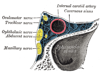


The Sinuses Of The Dura Mater Human Anatomy
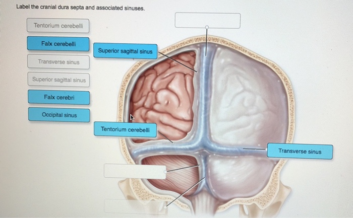


Label The Cranial Dura Septa And Associated Sinuses Chegg Com
Dural sinus stenting for pseudotumor cerebri was performed under general anesthesia Measurements of target sinus diameter and stenosis lengths were made from the initial diagnostic venogram All patients were premedicated with dual antiplatelet therapy A 6 or 8 Fr Pinnacle sheath was placed into the right common femoral vein utilizing aID Title Dural Venous Sinuses Category LabeledAnatomy Atlas 6E ID Category LabeledCochard Imaging 1E Radiographs ID Title Dural Venous Sinuses Category LabeledHansen FC 3E1219 · Dural venous sinuses Sural venous sinuses Source Medbullets At certain sites, the two layers of dura mater separate to form the dural venous sinuses – the system for venous drainage of the cranium and brain These are lined by vascular endothelium, with no valves or muscular tissue, and ultimately drain into the internal jugular veins


Groove For Sigmoid Sinus
:background_color(FFFFFF):format(jpeg)/images/article/en/sigmoid-sinus/ROdsfVByutGadvrRpHR9gQ_v1iO4kKNjZkWc9nKmI6Q_Sinus_sigmoideus_01.png)


Sigmoid Sinus Anatomy Kenhub
Perdense dural sinus to one that is isodense with brain) 7, 9 (Fig 4) Another cause is volume averaging of a dural sinus with normal tissue, which is most commonly encountered in the transverse sinus on axial CT images Fi nally, a small dural sinus, even when hyper dense, can be difficult to visualize against theThe contents of the dural venous sinuses ultimately drain into the internal jugular vein The dural venous sinuses can be either paired or unpaired Paired sinuses include the transverse sinus, cavernous sinus, superior petrosal sinus, inferior petrosal sinus, sphenoparietal sinus, and sigmoid sinus On the other hand, unpaired sinuses includeScience Quiz / Dural venous sinuses Random Science Quiz Can you name the Dural venous sinuses by Anatomy6511 Plays Quiz not verified by Sporcle Rate 5 stars Rate 4 stars Rate 3 stars Rate 2 stars Rate 1 star Forced Order Support Sporcle Go Orange Get the adfree and most optimal, fullfeatured Sporcle experience
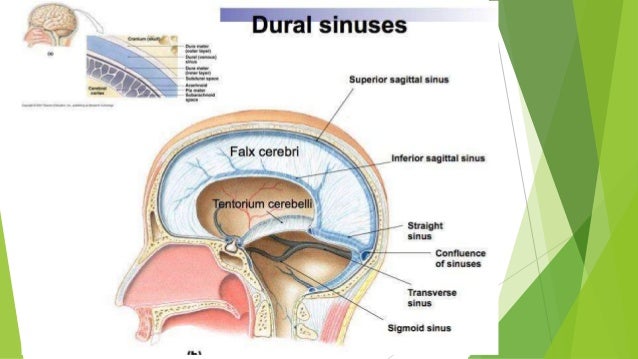


Dural Folds And Cavernous Sinus


Dural Venous Sinuses
· Dural Venous Sinuses Of The Brain Diagram Dural Venous Sinuses Of The Brain Diagram In this image, you will find superior sagittal sinus, falx cerebri, inferior sagittal sinus, straight sinus, cavernous sinus, transverse sinuses, sigmoid sinus, jugular foramen, right internal jugular vein in it Our SECOND youtube film is ready to runDural sinuses vary in size and shape in many pathological conditions with abnormal intracranial pressure Size and shape normograms of dural brain sinuses are not available The creation of such normograms may enable computerassisted comparison0109 · Dural sinus and cortical venous reflux (CVR), where the intravascular magnetically labeled proton is stagnant, are demonstrated as hyperintense signals at 15 s Slowflowing CVRs with prolonged ATT, which are not visible at 15 s, can be detected at 25 s


Dural Venous Sinuses 3d Anatomy Tutorial Youtube


Dural Venous Sinuses Wikipedia
Dural Arterivenous Fistula at the Cavernous Sinus Diagnosed by Arterial Spinlabeled Imaging Nobuaki Yamamoto, Yuki Yamamoto, Yuishin Izumi and Ryuji Kaji Abstract We herein report a case of dural arteriovenous fistula (DAVF) at the cavernous sinus that was diagnosed by arterial spinlabeled imaging (ASL) · Dural Venous Sinuses 1 Superior sagittal sinus lies at the superior attached border of falx cerebri Receives blood from superior cerebral veins (bridging veins) and emissary veins (connects extracranial venous system with intracranial venous sinuses · Dural venous sinuses Sagittal sinuses There are two sagittal sinuses that occupy the longitudinal cerebral fissure (midline between the Straight sinus Each anterior cerebral vein leaves the longitudinal cerebral fissure inferiorly and


Untitled Document


Venous Sinuses Of The Brain Google Search Con Imagenes Anatomia Medica
Online quiz to learn Dural venous sinuses; · The dural venous sinuses lie between the periosteal and meningeal layers of the dura mater They are best thought of as collecting pools of blood, which drain the central nervous system, the face, and the scalp All the dural venous sinuses ultimately drain into the internal jugular vein Unlike most veins of the body, the dural venous sinusesDemonstration of Dural Sinus Occlusion by the Use of MR Angiography David J Rippe,' Orest B Boyko,' Charles E Spritzer,1 William J Meisler,' Charles L Dumoulin,2 Steve P Souza,3 and E Ralph Heinz' · Thrombosis involving the dural venous sinuses is a well known clinical entity Patients present with a variety of symp


Superior Sagittal Sinus Location Function Human Anatomy Kenhub Youtube


Dural Venous Sinuses Of The Brain Diagram Quizlet
Dural venous channels — venous sinuses in the tentorium cerebelli that connect cerebellar and inferior cortical veins to the named venous sinuses (transverse, sigmoid) are well known There is surgical literature on being careful not to cut them and risk venous infarctionPulsatile Tinnitus of Dural Sinus Origin Dr Jackler and Ms Gralapp retain copyright for all of their original illustrations which appear in this online atlas We encourage use of our illustrations for educational purposes, but copyright permission should be sought before publication orTransverse dural sinuses Incidence of anatomical variants and flow artefacts with 2D timeofflight MR venography at 1 tesla Localization of Anterosuperior Point of Transversesigmoid Sinus Junction Using a Reference Coordinate System on Lateral Skull Surface


Q Tbn And9gctvzqget8dkmwqjtokuxc0bghdett2jtipdlppul 0 Usqp Cau


Brain
Today's Rank0 Today 's Points One of us! · Collectively, our results highlight the dural sinuses as a critical interface for immunebloodbrain interactions At these unique sites, all the components necessary for CNSantigen exposure, uptake by APCs, and presentation to patrolling dural T cells are functional, enabling immune surveillance of CNSantigensThey also drain CSF They communicate with veins outside the cranial cavity via emissary veins Name


Dural Venous Sinuses Radiology Reference Article Radiopaedia Org
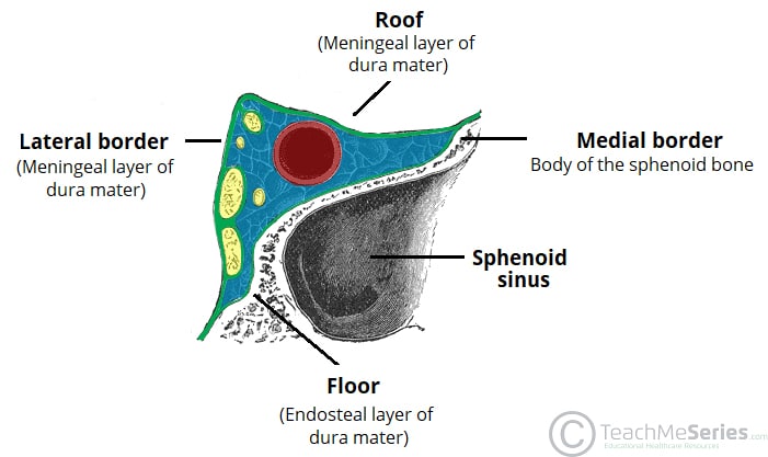


The Cavernous Sinus Contents Borders Thrombosis Teachmeanatomy
Rustenhoven et al identify the dural sinuses as a neuroimmune interface, where patrolling T cells survey brain and CSFderived antigens to enable CNS immune surveillance This niche is altered during aging and neuroinflammation and may represent a new therapeutic target forThe transverse sinuses, within the human head, are two areas beneath the brain which allow blood to drain from the back of the head They run laterally in a groove along the interior surface of the occipital bone They drain from the confluence of sinuses to the sigmoid sinuses, which ultimately connect to the internal jugular vein See diagram labeled under the brain as "SIN TRANS" The transverse sinuses · This clearly demonstrates that the dural sinuses are a major site of T cell surveillance from the blood stream


Dural Venous Sinuses
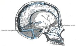


Confluence Of Sinuses Wikipedia
· Dural sinuses 1 Pronograde Quadruped vertebrate Orthograde – plantigrade Biped – erect vertebrate 2 Splanchnocranium Neurocranium Telencephalisation Inversely proportional 3 Blood supply of brain Venous drainage of brain Brain – Internal carotid Vertebral art Choroid plexus Skull – MidMeningeal art• ultimately drain into dural venous sinuses!• sits against lateral aspect of body of sphenoid!


Dural Venous Sinuses Diagram Quizlet


Temporal Bone Cranial Nerves And Dural Sinuses Labeled Download Scientific Diagram
· Few reports have addressed the surgical management of cranial metastases that overlie or invade the dural venous sinuses To examine the role of surgery in the treatment of dural sinus calvarial metastases, we reviewed retrospectively 13 patients who were treated with surgery at the University of Texas MD Anderson Cancer Center between 1993 and 1999Your Skills & Rank Total Points 0 Get started! · The dural venous sinuses (also called dural sinuses, cerebral sinuses, or cranial sinuses) are venous channels found between the endosteal and meningeal layers of dura mater in the brain It means that you have a gross patent and need a new one


The Sinuses Of The Dura Mater Human Anatomy
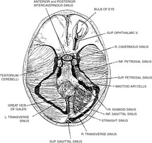


Venous And Dural Sinus Thrombosis Chapter 13 Vertebrobasilar Ischemia And Hemorrhage
• also emissary veins from outside the cranial cavity!!!!0516 · Dural sinus malformations (DSM) are rare pediatric vascular malformation Two types of DSMs have been recognized a midline type involving the posterior sinus with giant dural lakes and slow flow mural arteriovenous shunting and a lateral type involving the jugular bulb with otherwise normal sinuses and associated highflow sigmoid sinus arteriovenous fistulaWe herein report a case of dural arteriovenous fistula (DAVF) at the cavernous sinus that was diagnosed by arterial spinlabeled imaging (ASL) A 67yearold woman was referred to our hospital due to double vision and bilateral conjunctival injection Conventional magnetic resonance imaging findings were normal


Chapter 2 Part 6 Dural Sinuses And Veins Flashcards Quizlet
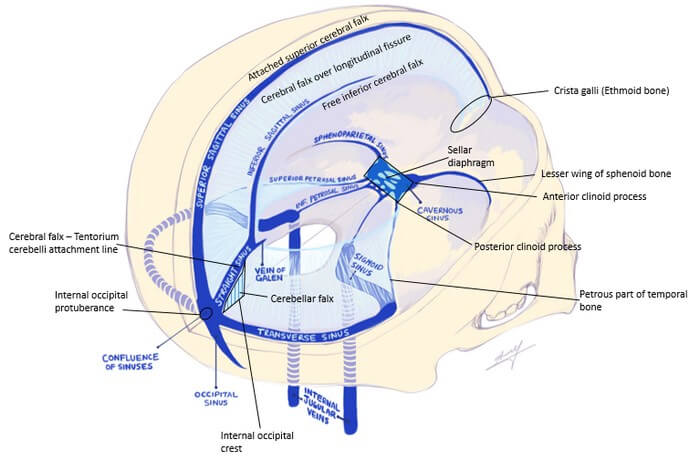


Dural Reflections And Venous Sinuses Epomedicine
· We herein report a case of dural arteriovenous fistula (DAVF) at the cavernous sinus that was diagnosed by arterial spinlabeled imaging (ASL) A 67yearold woman was referred to our hospital due to double vision and bilateral conjunctival injection Conventional magnetic resonance imaging findings were normalDural venous sinuses There are seven paired (transverse, cavernous, greater & lesser petrosal, sphenoparietal, sigmoid and basilar) and five unpaired (superior & inferior sagittal, straight, occipital and intercavernous) dural sinuses There are two sagittal sinuses that occupy the longitudinal cerebral fissure (midline between the cerebralThis video provides a walkthrough of the dural venous sinuses (eg transverse sinus, cavernous sinus) You can follow along using our free written guide wit


Dural Sinuses


1
ID Title Venous Sinuses Category LabeledFelten 2E ID 523 Title Venous Sinuses Category LabeledFelten Flash Cards 2E ID Title Venous Sinuses Sinus DurThe dural venous sinuses (also called dural sinuses, cerebral sinuses, or cranial sinuses) are venous channels found between the endosteal and meningeal layers of dura mater in the brain 1 2 They receive blood from the cerebral veins , receive cerebrospinal fluid (CSF) from the subarachnoid space via arachnoid granulations , and mainly empty into the internal jugular veinDural sinus Any of several large endothelialined collecting channels into which veins of the brain and inner skull empty and which then empty into the internal jugular vein These venous sinuses are found between the two layers (periosteal and meningeal) of the dura mater Their walls have no muscle, and they have no valves to give direction


Cerebral Circulation And Metabolism Sciencedirect
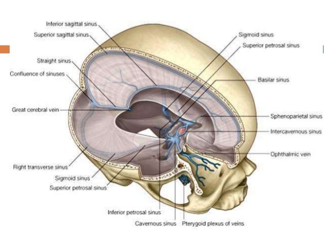


15 Dural Venous Sinuses
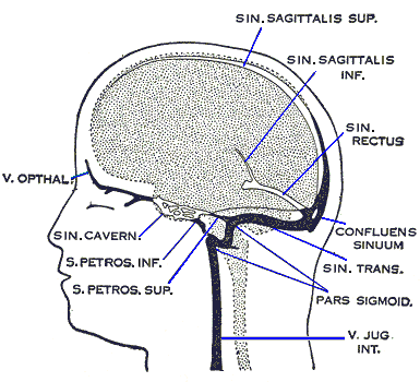


Anatomy And Function Of The Dural Venous Sinuses Medical Library
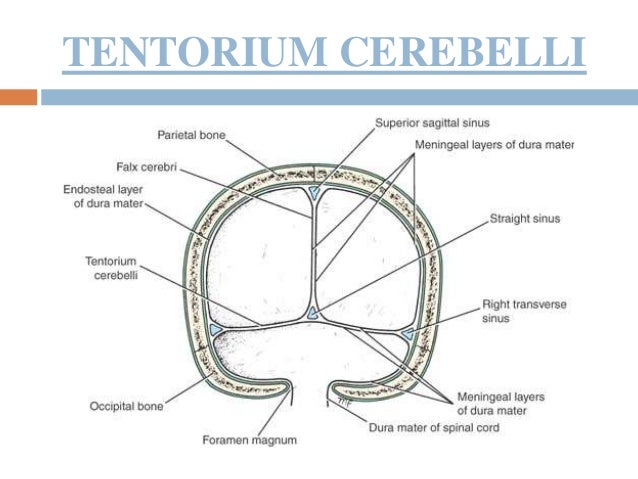


15 Dural Venous Sinuses
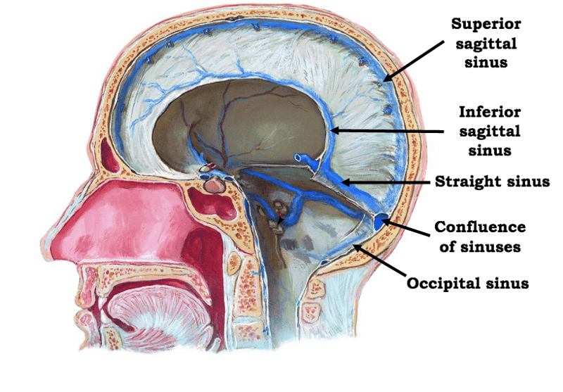


Ventricles Meninges Csf And Blood Vessels Of The Brain Flashcards Easy Notecards


Dural Sinuses


Dural Venous Sinuses Anatomy Anatomy Drawing Diagram
:background_color(FFFFFF):format(jpeg)/images/article/en/dural-sinuses/MBT9Z5mR2o8TnY2hb131w_Superficial_veins_of_the_brain_lateral_view.png)


Dural Venous Sinuses Anatomy Kenhub


Dural Venous Sinus Anatomy
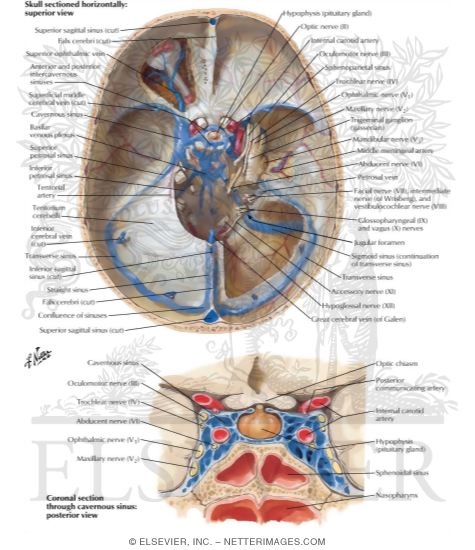


Dural Venous Sinuses


The Sinuses Of The Dura Mater Human Anatomy


Straight Sinus Ultrastructural Analysis Aimed At Surgical Tumor Resection In Journal Of Neurosurgery Volume 125 Issue 2 16


Grooves For Dural Venous Sinuses Paranasal Sinuses Diagram Quizlet


Dural Venous Sinuses Radiology Reference Article Radiopaedia Org


Mrv Major Venous Sinuses Are Labeled With Arrows Download Scientific Diagram


Temporal Bone Cranial Nerves And Dural Sinuses Labeled Download Scientific Diagram


Cerebral And Sinus Vein Thrombosis Circulation


Venous Sinuses Neuroangio Org
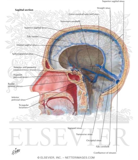


Dural Venous Sinuses
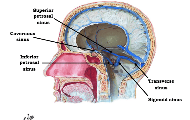


Print Ventricles Meninges Csf And Blood Vessels Of The Brain Flashcards Easy Notecards
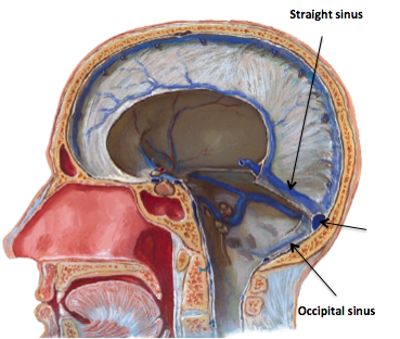


Dural Venous Sinuses Flashcards Memorang
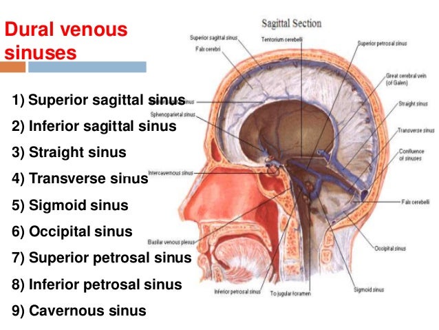


15 Dural Venous Sinuses


Dural Venous Sinuses Diagram Quizlet


Http Www Headandnecktrauma Org Wp Content Uploads 16 10 Meninges And Dural Sinuses Pdf


Dural Venous Sinuses And Veins Of Head And Neck Labeled Eccles Health Sciences Library J Willard Marriott Digital Library
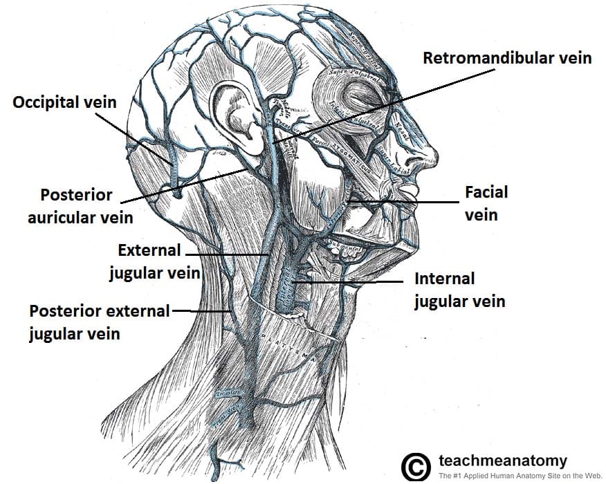


Venous Drainage Of The Head And Neck Dural Sinuses Teachmeanatomy


Dural Venous Sinuses Ventricles Diagram Quizlet


Dural Sinuses
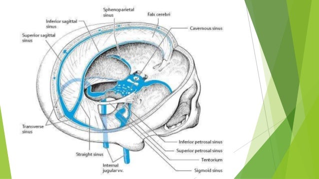


Dural Folds And Cavernous Sinus


Transverse Sinuses Wikipedia
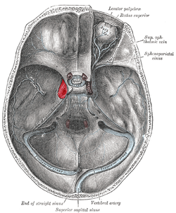


Cavernous Sinus Wikipedia
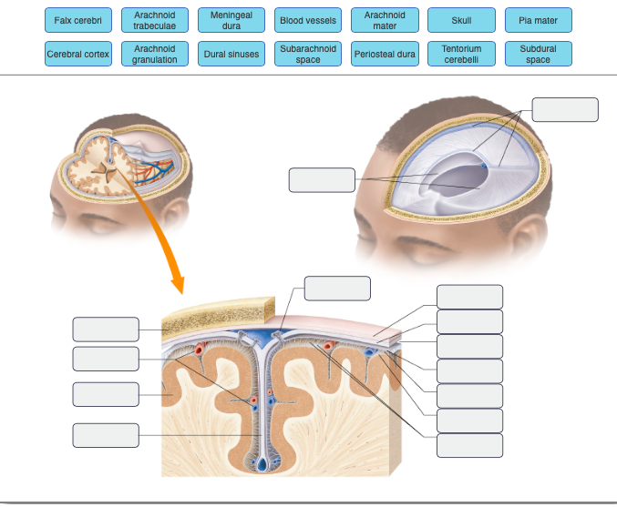


Meningealblood Vessels Arachnoid Mater Arachnoid Chegg Com


Venous Drainage Of The Brain Anatomy Geeky Medics


1


Dural Venous Sinuses Wikipedia
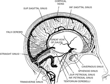


Venous And Dural Sinus Thrombosis Chapter 13 Vertebrobasilar Ischemia And Hemorrhage


Dural Sinuses Diagram Quizlet


Final Anatomy Brain Cranial Nerves 15b Flashcards Quizlet
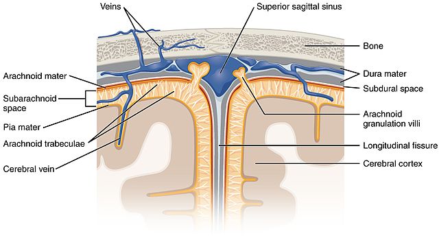


Anatomy And Function Of The Dural Venous Sinuses Medical Library


Dural Venous Sinuses
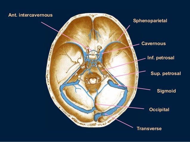


Dural Sinuses
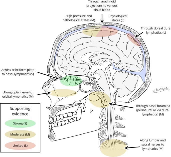


Cerebrospinal Fluid Outflow A Review Of The Historical And Contemporary Evidence For Arachnoid Villi Perineural Routes And Dural Lymphatics Springerlink
:background_color(FFFFFF):format(jpeg)/images/library/13675/meninges-and-arachnoid-granulations_english.jpg)


Superior Sagittal Sinus Anatomy Tributaries Drainage Kenhub
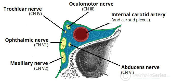


Venous Drainage Of The Head And Neck Dural Sinuses Teachmeanatomy


Cambridge Questions


1
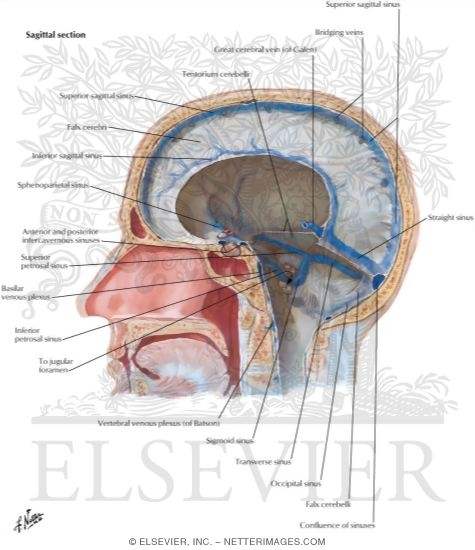


Dural Venous Sinuses


Dural Venous Sinuses Of The Brain Diagram
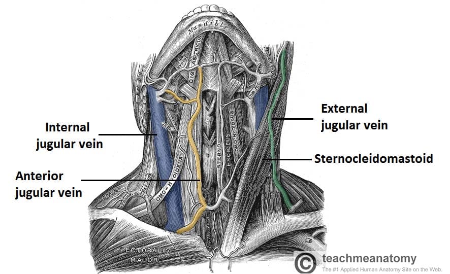


Venous Drainage Of The Head And Neck Dural Sinuses Teachmeanatomy
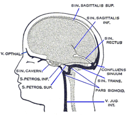


Superior Sagittal Sinus Wikipedia
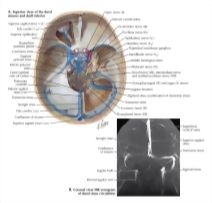


Dural Venous Sinuses
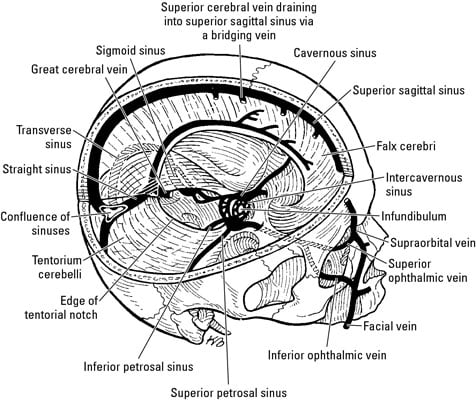


Anatomy Of The Brain The Meninges Dummies


Pin On Brain


Venous Sinuses Neuroangio Org


Dural Sinuses


Pin On Cns


File Dural Sinuses Jpg Wikimedia Commons


Venous Sinuses Neuroangio Org
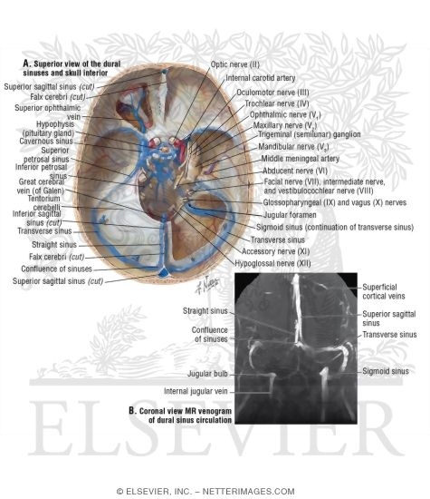


Dural Venous Sinuses
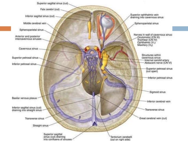


15 Dural Venous Sinuses


Untitled Document
:background_color(FFFFFF):format(jpeg)/images/article/en/superior-sagittal-sinus/ypAElOcFaTzHHpIP8R3qw_8iVwiSwwNxvJhpSQELoQ_Sinus_sagittalis_superior_02.png)


Superior Sagittal Sinus Anatomy Tributaries Drainage Kenhub


Http Www Headandnecktrauma Org Wp Content Uploads 16 10 Meninges And Dural Sinuses Pdf
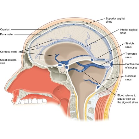


Dural Venous Sinuses Radiology Reference Article Radiopaedia Org


Cerebral And Sinus Vein Thrombosis Circulation


Dural Venous Sinuses
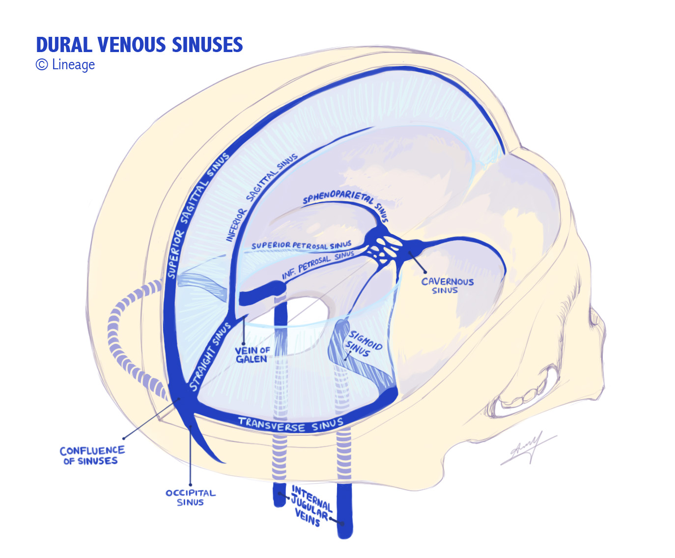


Dural Venous Sinuses Medmule


Functional Characterization Of The Dural Sinuses As A Neuroimmune Interface Sciencedirect


Dural Venous Sinuses Anatomy Kenhub


Pin On Nbde Part I
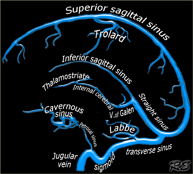


The Radiology Assistant Cerebral Venous Thrombosis


Cavernous Sinus Anatomy Anatomy Drawing Diagram


Venous Sinuses Neuroangio Org
:background_color(FFFFFF):format(jpeg)/images/library/11573/the-dural-venous-sinuses_english.jpg)


Meninges Ventricles Csf And Brain Blood Supply Kenhub
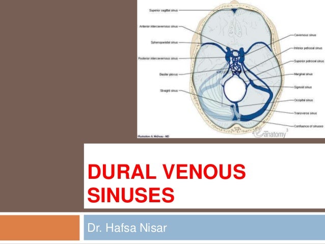


15 Dural Venous Sinuses


Pin On Ot Neuroscience


Dural Sinuses
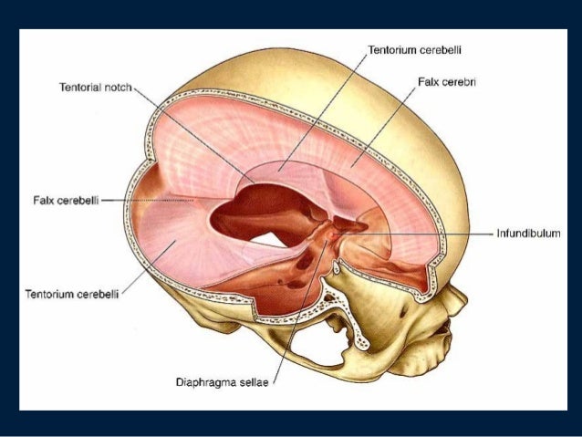


Dural Sinuses


コメント
コメントを投稿